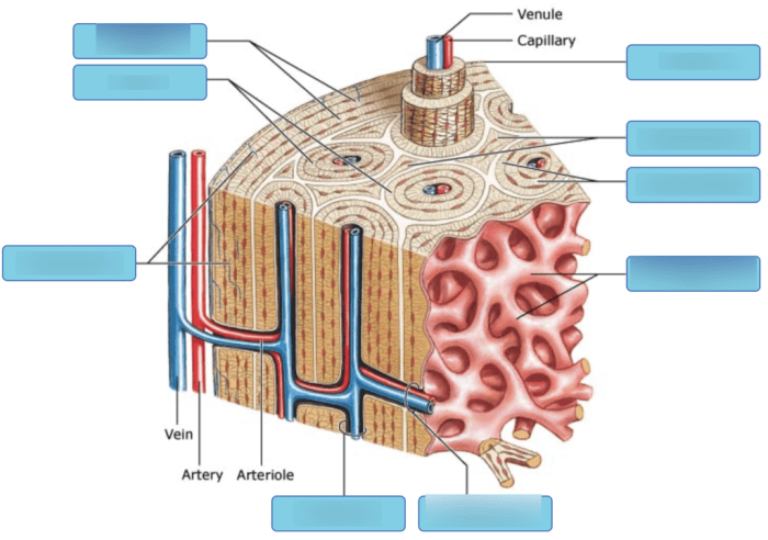
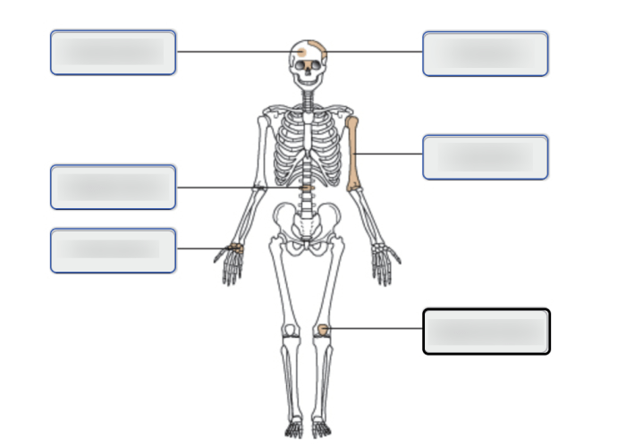
Art-labeling activity structure of a long bone provides a comprehensive overview of the essential components of a long bone, offering a detailed examination of its anatomy and the significance of accurate labeling. This guide delves into the educational and clinical applications of art-labeling, exploring advanced techniques and their potential impact on bone anatomy education and research.
Bone Anatomy

Long bones are the primary structural components of the human skeleton. They are characterized by their elongated, cylindrical shape and are responsible for providing support, protection, and movement.
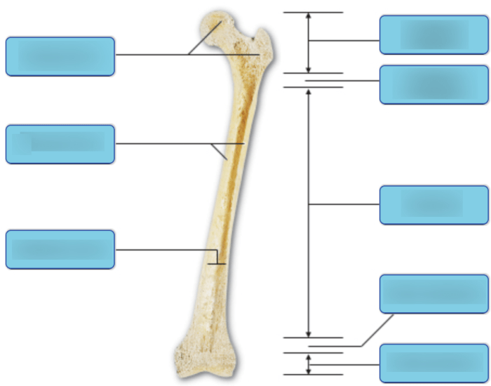
The general structure of a long bone consists of three main regions:
- Diaphysis:The shaft of the bone, which is the main cylindrical portion.
- Epiphysis:The expanded ends of the bone, which articulate with other bones at joints.
- Metaphysis:The transitional zone between the diaphysis and epiphysis, where growth occurs during childhood.
The outer surface of the bone is covered by a thin membrane called the periosteum, which provides nourishment and protection. The inner surface of the bone is lined by the endosteum, which is responsible for bone remodeling.
Art-Labeling Activity

Art-labeling activities are a valuable educational tool for enhancing understanding of bone anatomy. By labeling the different anatomical landmarks on a long bone, students can reinforce their knowledge and develop a deeper appreciation for the complexity of the skeletal system.
Here are some specific anatomical landmarks that can be labeled on a long bone art:
- Diaphysis
- Epiphysis
- Metaphysis
- Periosteum
- Endosteum
- Medullary cavity
- Nutrient foramen
- Articular cartilage
Accurate labeling is crucial for ensuring that students develop a clear understanding of the bone’s anatomy. It allows them to identify and locate specific structures, which is essential for both educational and clinical applications.
Structure of the Art

The typical arrangement of labels on a long bone art follows a logical and consistent pattern.
- The diaphysis is typically labeled along its length, with the proximal end (closest to the body) labeled first.
- The epiphyses are labeled separately, with the proximal epiphysis labeled first, followed by the distal epiphysis.
- The metaphysis is labeled as the transitional zone between the diaphysis and epiphysis.
- Other anatomical landmarks are labeled in relation to their position on the bone.
Colors, lines, and symbols are often used to enhance the clarity and effectiveness of labeling. For example, different colors may be used to differentiate between different bone regions, while lines and arrows may be used to indicate the direction of structures.
Variations in Labeling: Art-labeling Activity Structure Of A Long Bone

While there is a general consensus on the labeling of long bone anatomy, there may be some variations in conventions across different sources.
These variations can arise due to:
- Differences in terminology
- Variations in the level of detail included
- Differences in the intended audience
Despite these variations, it is important to maintain consistency in labeling to ensure clarity and avoid confusion.
Q&A
What is the importance of accurate labeling in art-labeling activity?
Accurate labeling is crucial for conveying precise anatomical information and ensuring consistent communication among healthcare professionals and educators.
How can art-labeling activities enhance understanding of bone anatomy?
Art-labeling activities actively engage students in the learning process, promoting a deeper understanding of bone anatomy through visual representation and hands-on labeling.